 >
>
Concept Graph & Resume using Claude 3 Opus | Chat GPT4 | Llama 3:
Resume:
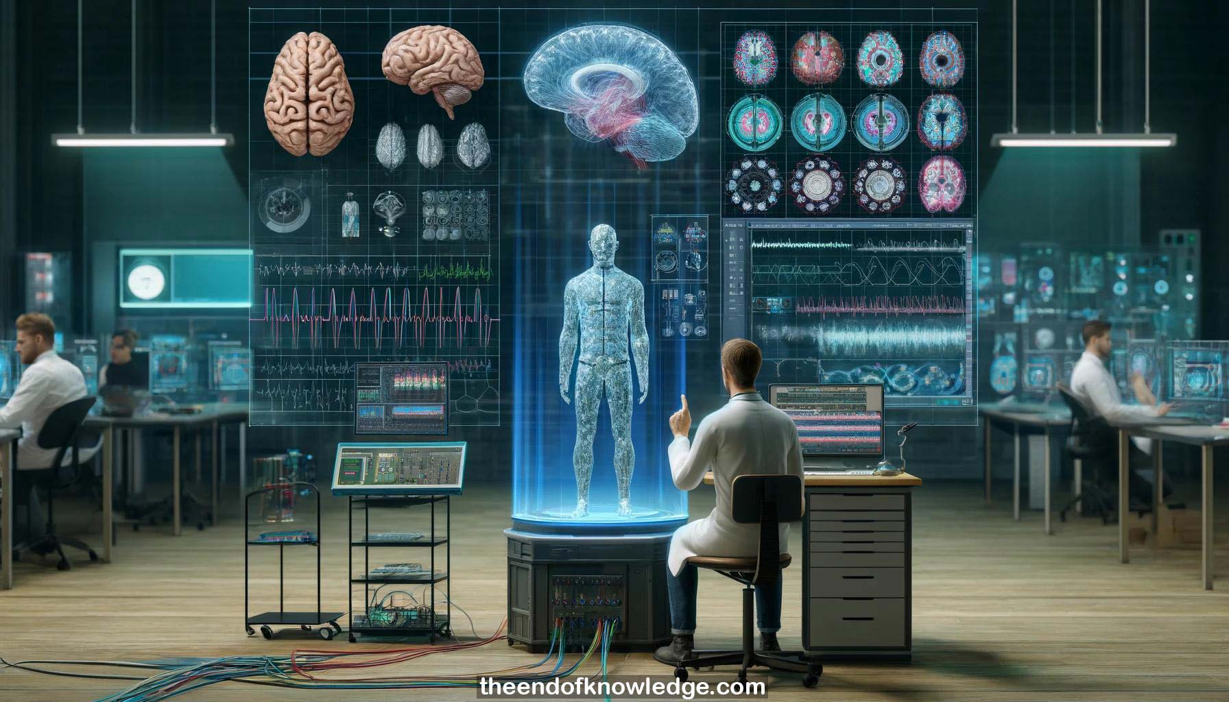
1.- Michael Jordan, a skilled programmer, presents on 3D mapping of grid and stereo EEG activity and high frequency oscillation detection.
2.- Defining electrode montages representing signal locations is important for topographical visualizations, anatomical mapping, and comparisons to other modalities like fMRI.
3.- Simple 2D montages use schematic images or photos, while 3D montages use template brains or subject-specific MRI-based brain models.
4.- The IAG Montage Creator is a standalone application that creates 2D and 3D montages exportable as a single file.
5.- It imports data from FreeSurfer, a powerful free tool that reconstructs 3D brain models from MRI scans in 5-24 hours.
6.- It also imports cortex surfaces from other tools, resection areas, DTI tracks, and allows manual electrode placement aided by parcellation maps.
7.- Post-operative CT scans enable automatic localization of grid and depth electrodes, compensating for brain shift between pre- and post-operative scans.
8.- Coordinates from other systems can be imported to semi-automatically place electrodes on the 3D model.
9.- The montages are used for real-time 3D visualization in MATLAB/Simulink and offline plotting for analysis and publication.
10.- Mostafa Mohammadpour discusses epilepsy biomarkers - the seizure onset zone, irritative zone of interictal spikes, and high frequency oscillations (HFOs).
11.- Seizures show initiation, increase in frequency/amplitude, and termination patterns. The seizure onset zone is a "gold standard" for surgical resection.
12.- Spikes last 20-200ms. Pathological HFOs >80Hz riding on spikes can localize the seizure onset zone better than spikes alone.
13.- Challenges in HFO detection include artifacts. True HFOs appear as "islands" in time-frequency maps while artifacts elongate across frequencies.
14.- Existing time-domain HFO detectors bandpass filter, calculate energy, threshold, and apply duration and peak number criteria.
15.- The authors aimed to automatically detect seizure onset zones, which are usually marked subjectively by doctors after reviewing multiple seizures.
16.- They collected a dataset of several seizure onset patterns, extracted time and time-frequency features, and trained an LDA classifier.
17.- Classification performance was good overall but weaker for uncommon onset patterns like delta rhythms due to imbalanced data.
18.- Next, they detected sequences of propagating spikes across adjacent electrodes within 15-50ms and 2-5cm using an existing algorithm.
19.- To identify true spikes, they clustered detected spikes by shape using DTW distance and kept clusters resembling textbook spike morphologies.
20.- 35% of true spikes and 62% of non-spikes localized to seizure onset zones. True spikes clustered closer to onset zones.
21.- In 3 patients, they compared rates of spikes, ripples (80-250Hz), fast ripples (250-500Hz), and pathological HFOs between medicated and unmedicated states.
22.- Event rates were higher off medication as expected. Optimizing rate thresholds yielded 95% specificity for localizing seizure onset zones.
23.- Spike sequences and pathological HFOs localized closest to onset zones, outperforming isolated spikes, ripples and fast ripples.
24.- Lastly, they developed a real-time Simulink model to detect spikes using adaptive thresholds that stabilize after ~3 minutes of data.
25.- The real-time detector performed comparably to offline methods. Spikes were visualized on a 3D brain and as per-minute spike rates.
Knowledge Vault built byDavid Vivancos 2024