 >
>
Concept Graph & Resume using Claude 3 Opus | Chat GPT4 | Llama 3:
Resume:
1.-Christoph Kapeller leads G-Tech's invasive department, working on cortical technologies, intracranial electrical stimulation, and neuromodulation experiments using ECoG implants.
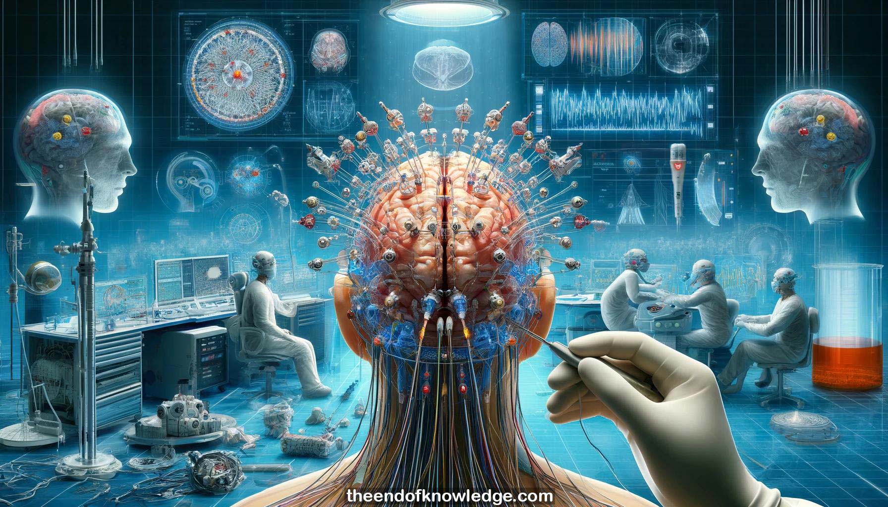
2.-ECoG grids with 20-64 channels are implanted on cortical regions by neurosurgeons to record high gamma and other oscillations up to 1kHz.
3.-Depth electrodes (stereo EEG) can be inserted into the brain to record from deep structures, in addition to ECoG.
4.-ECoG has higher amplitude, frequency range, and spatial resolution compared to EEG. Stereo EEG and ECoG have similar electrode properties.
5.-ECoG grids are placed subdurally during craniotomy. Stereo EEG electrodes are implanted through screw holes without removing the skull.
6.-Electrodes have passive connections to a head box which connects to the biosignal amplifier. Quick connectors can interface multiple electrodes.
7.-Intracranial signals have stronger amplitude and higher frequency range than EEG. ECoG and stereo EEG can record up to 1kHz.
8.-High gamma band shows focused cortical activation during tasks, while low frequencies show widespread suppression. High gamma relates to neural firing.
9.-ECoG cursor control, video game interfaces, and robotic control have been demonstrated using motor cortex signals in the past.
10.-Clinical opportunity, limited time, protocol design, channel assignment, control computer, processing platform, and analysis pipeline are key experiment considerations.
11.-Ground and reference electrode selection, connection to earth ground, and proper line noise management are crucial for good signal quality.
12.-Impedance checking helps identify bad channels. Operating rooms introduce more line noise interference than monitoring units. Proper referencing helps.
13.-Sampling rates of 1200Hz and higher are used. 24-bit ADCs allow wide input range. Passive cable connections go to the amplifier.
14.-Experiment design uses an XML-based task protocol loaded into GHYSIS/Simulink. Raw data visualization and online processing are set up.
15.-Offline data review in MATLAB with GBS Analyze checks signal quality, re-references data, and defines task-related analysis intervals.
16.-Time-frequency analysis from 5-145Hz and topographic mapping to grid layouts help visualize task-related high gamma biomarkers across the grid.
17.-Classifier training data is extracted, spatially filtered with CSP, band-power extracted, and used to train an LDA classifier.
18.-Online BCI output is generated by spatially and temporally filtering data, extracting normalized high gamma band-power, and classifying it.
19.-The real-time BCI is validated with the subject, showing fast decoding of presented faces vs symbols from untrained images.
20.-The same signal processing pipeline is applied for continuous decoding for robotic arm control based on motor cortex activity.
21.-Anatomical localization of electrodes on the brain is done by co-registering pre-op MRI, post-op CT and segmenting the brain.
22.-Electrodes are snapped to the brain surface segmentation to correct for brain shift. Stereo EEG electrodes are localized along shafts.
23.-3D coordinates of electrodes are determined and assigned to brain regions. fMRI co-registration can map function and symptoms.
24.-Group-level anatomical localization on template brains allows mapping biomarker and symptom topology across subjects with variable grid locations.
25.-Effective stimulation mapping and passive BCI experiments are enabled. Diagnostic functional mapping is an important use case.
26.-Impedance and epileptic activity can also be visualized spatially. Connectivity analysis can be applied using BCIs.
27.-The core pipeline is ECoG/stereo EEG recording, spatial and temporal filtering, normalization, classification, and output generation for real-time operation.
28.-Careful design of protocols, signal processing and validation allows for effective ECoG BCIs from the operating room to chronic implants.
29.-Electrode localization and integration with other brain mapping modalities helps interpret mechanisms and localize function for many use cases.
30.-Despite penetrating the brain, stereo EEG appears generally safe other than bleeding risks. Symptoms specifically from the shafts are not obvious.
Knowledge Vault built byDavid Vivancos 2024