 >
>
Concept Graph & Resume using Claude 3 Opus | Chat GPT4 | Llama 3:
Resume:
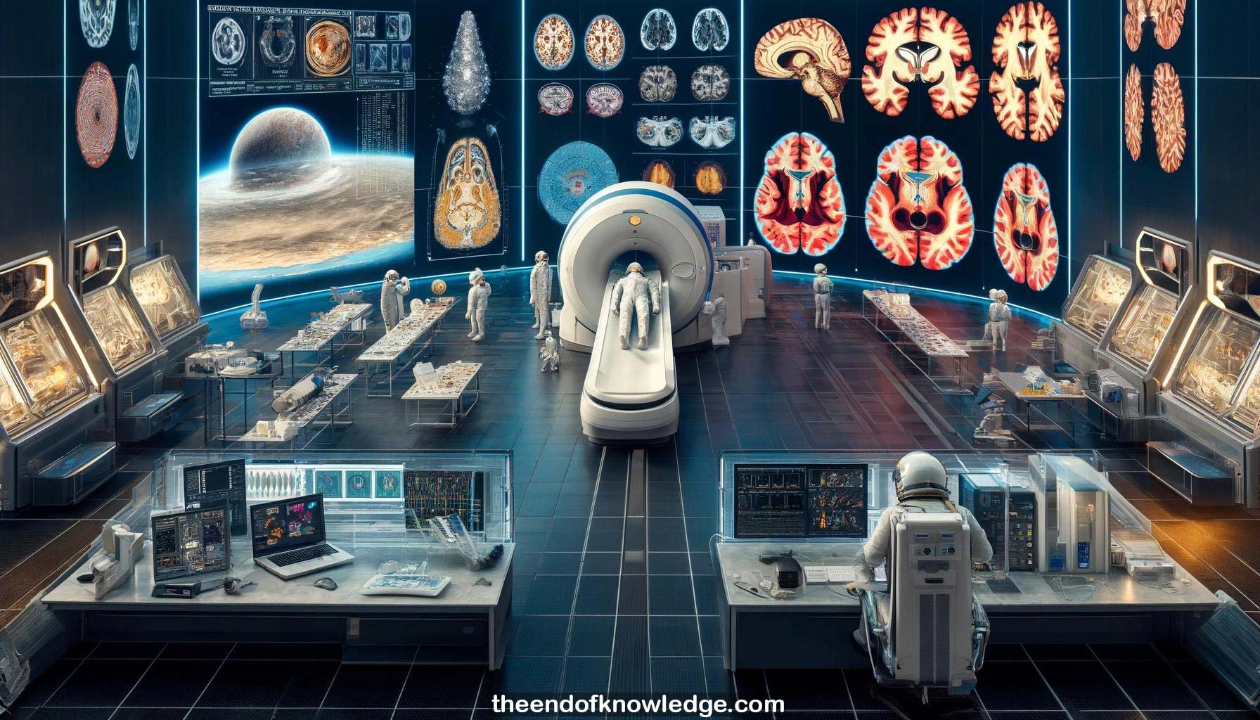
1.- Steven Jillings from University of Antwerp studies changes in astronauts' brains due to microgravity using MRI scans before and after spaceflight.
2.- The human body, including the vestibular system and brain, has evolved under constant 1G gravity and is affected by microgravity.
3.- Studying astronaut brains helps monitor changes from the hostile space environment, including radiation, microgravity, isolation, and high workload.
4.- The vestibular system, which includes otoliths in the inner ear, senses gravity and head movements. It integrates with visual and proprioceptive systems.
5.- Neuroplasticity is the brain's ability to change structure and function in response to development, learning, environment, and spaceflight.
6.- Their longitudinal MRI study scans astronauts before spaceflight, immediately after return, and 6 months later, using anatomical, diffusion, and functional MRI.
7.- Initial anatomical MRI analysis using voxel-based morphometry showed large decreases in gray matter volume in the temporal, parietal and frontal lobes post-flight.
8.- CSF volume decreased at the top of the brain and increased at the bottom and in ventricles, suggesting an upward brain shift.
9.- Gray matter volume changes were less extensive 6 months post-flight, indicating they are not atrophy but rather fluid shift related.
10.- Other research groups found similar evidence of brain tissue crowding at the top and narrowing of sulci, corroborating the upward brain shift.
11.- Ventricular volume increased over 12% immediately post-flight and remained 5% larger 6 months later, an effect size much larger than typical aging.
12.- Diffusion MRI analysis showed gray matter crowding at the top matched the volumetric findings, with no net gray matter tissue loss.
13.- CSF redistribution drives the brain position changes, with CSF decreasing at the top and increasing at the bottom of the brain.
14.- The brain shift effects partially resolve from immediate post-flight to 6 months later but some changes persist compared to pre-flight.
15.- Perivascular space volumes increased post-flight, were higher in astronauts with spaceflight-associated neuro-ocular syndrome (SANS), and differed between NASA and Roscosmos crews.
16.- Ventricular expansion may act as a buffer for intracranial fluid increases and was lower in SANS, suggesting a role in its pathophysiology.
17.- Differences between NASA and Roscosmos crews may relate to use of resistive exercise vs. lower body negative pressure as countermeasures, respectively.
18.- Brain shift modeling suggests it would enhance the effects of transcranial magnetic stimulation by reducing CSF space between the brain and stimulation site.
19.- In summary, the major fluid shift effects include gray matter volume decreases (but not atrophy), CSF redistribution, ventricular expansion, and perivascular space increases.
20.- Diffusion MRI also found signs of neuroplasticity, with localized white and gray matter tissue increases in the basal ganglia, cerebellum and motor cortex.
21.- Tractography analysis revealed changes in cerebellar, corticospinal, corticostriatal, callosal and arcuate fasciculus tracts, some related to motor function and others possibly to deformation.
22.- Fiber density and cross-section metrics confirm net tissue increases in the cerebellum's middle and superior peduncles, the clearest evidence of neuroplasticity.
23.- Functional MRI found altered connectivity in the posterior cingulate, thalamus, angular gyrus and insula, regions involved in environmental monitoring, cognition, and vestibular processing.
24.- The angular gyrus shows decreased connectivity after both spaceflight and parabolic flight and increased connectivity in fighter pilots, suggesting gravity-dependent neuroplasticity.
25.- Angular gyrus connectivity changes also correlate with working memory performance and vestibular function measures, further indicating its role in gravity adaptation.
26.- Planned parabolic flight studies will examine angular gyrus activity during a vertical oddball task using EEG to clarify its link to spatial perception.
27.- In conclusion, spaceflight induces fluid shifts and brain position changes as well as sensorimotor and vestibular neuroplasticity, with the angular gyrus as a key region.
28.- Some spaceflight-induced brain changes persist 6 months post-flight, so complete recovery remains unknown and long-term effects require further study.
29.- Future work aims to assess impacts of mission duration, correlations with performance and clinical outcomes, early recovery, and long-term cumulative effects.
30.- Defining biomarkers and effective countermeasures is crucial to monitor brain health during future long-duration missions, such as to Mars.
Knowledge Vault built byDavid Vivancos 2024